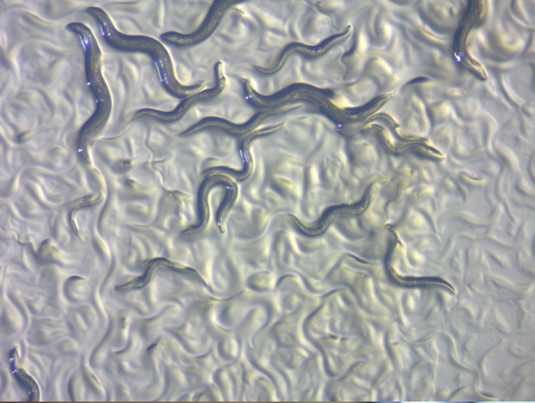
Princeton researchers have developed a way to track the activity of individual neurons while a worm is moving. Until recently, scientists have relied on methods that require the animal to be immobilized or that can only measure small sections of its brain.
However, in a recent study published in the Proceedings of the National Academy of Sciences (USA), physicists and neurobiologists have worked together to hone a calcium imaging technique that allows them to non-invasively record activity from all neurons in a worm’s brain while it is freely moving. This technique represents the first time that scientists have been able to record individual neuronal dynamics across the whole brain of an unrestrained animal.
The scientists used a small worm known as Caenorhabditis elegans or C. elegans. C. elegans is an ideal test system because of its compact nervous system of only 302 neurons that have already been comprehensively mapped. They genetically modified the worms to include two different types of dyes in each neuron in the brain. The first dye, red fluorescent protein (RFP), always glows red in the presence of an excitatory laser, which allowed the scientists to identify the position of each neuron that was visible in the setup.
The other dye, GCaMP6s, glows green in the presence of the laser, but only when there is an influx of calcium ions into the cell. An increase in calcium concentration in a neuron indicates that it is actively signaling, so the presence of both red and green fluorescence in a neuron meant that it was active.
By measuring the ratio of the always-present RFP fluorescence to the GCaMP6s fluorescence, researchers identified which neurons were firing at any given moment and correlated these to movement. They were able to map brain-wide neural activity to behaviors like moving forward, backward, turning or pausing.
In the future, these researchers hope to identify the neural activity behind an even wider range of behaviors, which will lead to a better understanding of how patterns in neural activity drive behavior, and could impact the understanding of human neurological disorders as well. Evidence has long suggested that many behaviors are driven by collective activity of many neurons, instead of just a few localized neurons, and this technique in small worms paves the way for scientists to explore these relationships.
Categories:
Tigra Scientifica: Whole Brain Imaging on the Go
Gabriella Wheeler, Contributor
October 31, 2018
0
Donate to The Tiger
Your donation will support the student journalists of Clemson University. Your contribution will allow us to purchase equipment and cover our annual website hosting costs.
More to Discover


